Chapter 1: Foundations of Biomedical Imaging: Principles, Modalities, and Emerging Technologies
1.1 The Role and Importance of Medical Imaging
Medical imaging plays a foundational role in modern healthcare by allowing clinicians to look inside the human body to diagnose, monitor, and treat disease. It transforms invisible biological processes into visible information, helping doctors detect conditions early, guide interventions, and evaluate how well treatments are working. From identifying a fractured bone to tracking the progression of cancer, medical imaging directly contributes to saving lives and improving patient outcomes.
Bioimaging, also called biomedical imaging or medical imaging, refers to a collection of technologies that create visual representations of structures and functions within the body. These terms are often used interchangeably and all describe the same core idea: using physics, engineering, and computation to visualize anatomy, physiology, and molecular activity. Each imaging technique is designed to highlight different features, offering complementary views of the same biological system.
As humans, we cannot see through solid objects. Medical imaging effectively gives us this ability—often compared to having “X-ray vision”—but without cutting into the body. Most imaging techniques are noninvasive, meaning they do not require surgical incisions. This dramatically reduces risk, pain, and recovery time for patients while still providing critical internal information.
The noninvasive nature of medical imaging is one of its greatest benefits in healthcare. It enables repeated measurements over time, supports preventive medicine, and allows clinicians to make informed decisions quickly and safely. In short, medical imaging bridges the gap between what we can observe externally and what is happening internally, making it an indispensable tool in modern medicine and a cornerstone of bioimaging research.
1.2 Discovery of X-Rays and the Origins of Medical Imaging
The field of medical imaging began in 1895 with the discovery of X-rays by Wilhelm Conrad Röntgen. While experimenting with cathode rays, Röntgen observed that an unknown form of radiation could pass through solid objects and expose photographic plates. He called this radiation “X-rays,” using X to indicate their unknown nature. This discovery marked a turning point in both physics and medicine, as it revealed a way to see inside the human body without surgery.
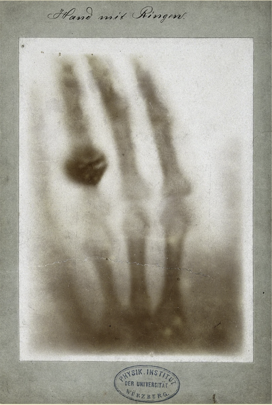
Figure 1. X-ray image of Wilhelm Conrad Röntgen's wife's hand, 1895. |
One of the most famous images in scientific history is the X-ray of Röntgen’s wife’s hand. The image clearly showed her bones and wedding ring, making the invisible visible for the first time. This striking demonstration immediately captured public and medical attention and showed the immense diagnostic potential of X-rays. Within just a few years, hospitals around the world began adopting X-ray imaging.
1.3 X-Ray Radiography
X-ray radiography is one of the simplest and most widely used medical imaging techniques. In this method, X-rays pass through the body and are absorbed differently by various tissues. Dense materials such as bone absorb more X-rays and appear bright on the image, while softer tissues absorb fewer X-rays and appear darker. This contrast makes X-ray radiography especially useful for detecting broken bones, joint problems, lung conditions, and certain tumors.
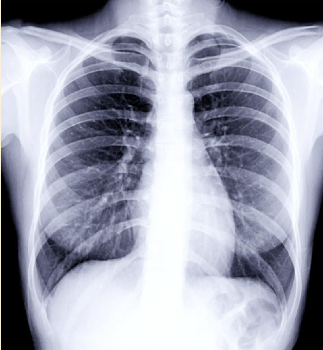
Figure 2. Black-and-white X-ray image showing a human ribcage. |
However, X-ray radiography has important limitations. The resulting image is two-dimensional, meaning that all structures along the path of the X-ray beam are compressed into a single flat image. As a result, overlapping tissues can obscure important details, making it difficult to clearly distinguish individual structures. This lack of depth information motivated the development of more advanced imaging techniques, such as computed tomography (CT), which can separate structures in three dimensions. Despite its limitations, X-ray radiography remains a cornerstone of medical imaging due to its speed, accessibility, and diagnostic value, and it laid the foundation for the entire field of bioimaging.
1.4 X-Ray Computed Tomography (CT)
While conventional X-ray radiography is fast and effective, it struggles when many anatomical structures are packed closely together. Because all tissues are projected onto a single 2D image, overlapping bones and organs can obscure important details. This limitation becomes especially problematic when imaging complex regions of the body such as the brain, chest, or abdomen.
X-ray computed tomography, commonly known as CT, was developed to overcome this challenge. The word tomography comes from the Greek words tomos (slice) and graphein (to write), meaning “imaging by slices.” Instead of producing one flat image, CT creates cross-sectional images—thin slices of the body—that reveal internal structures layer by layer.
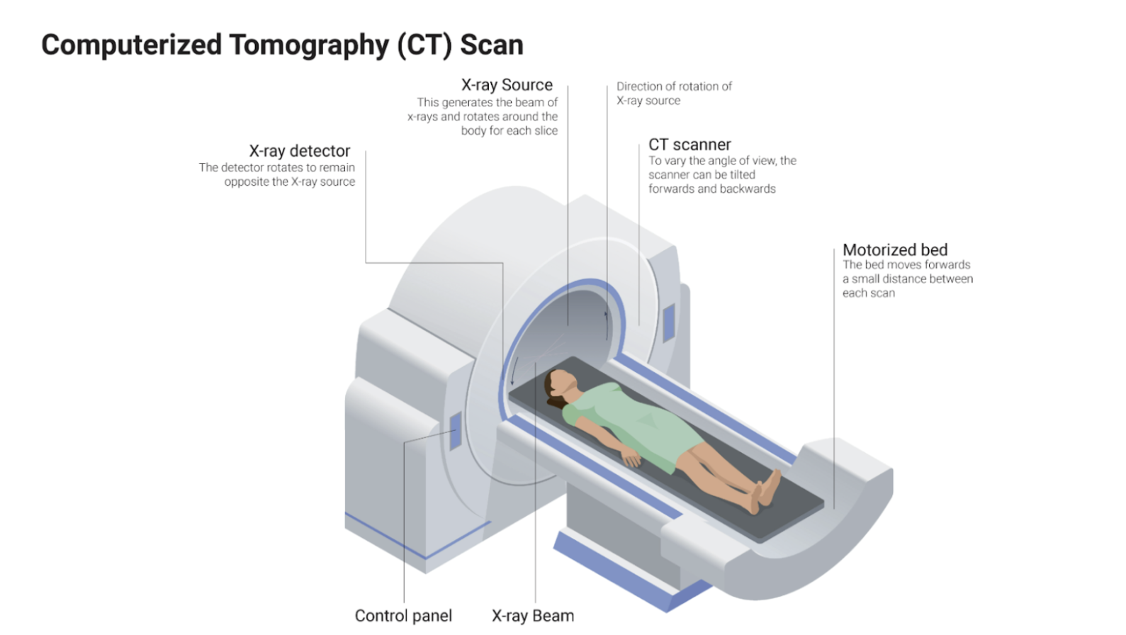
Figure 3. Diagram of a computerized tomography (CT) scanner showing key components. Source: [insert source here]. |
In a CT scan, an X-ray source and detector rotate around the patient, collecting projection images from many different angles. These multiple measurements capture how X-rays are absorbed along thousands of paths through the body. A computer then uses mathematical reconstruction algorithms to combine this information and generate a detailed image of a single slice. By stacking many slices together, clinicians can visualize the body in three dimensions.
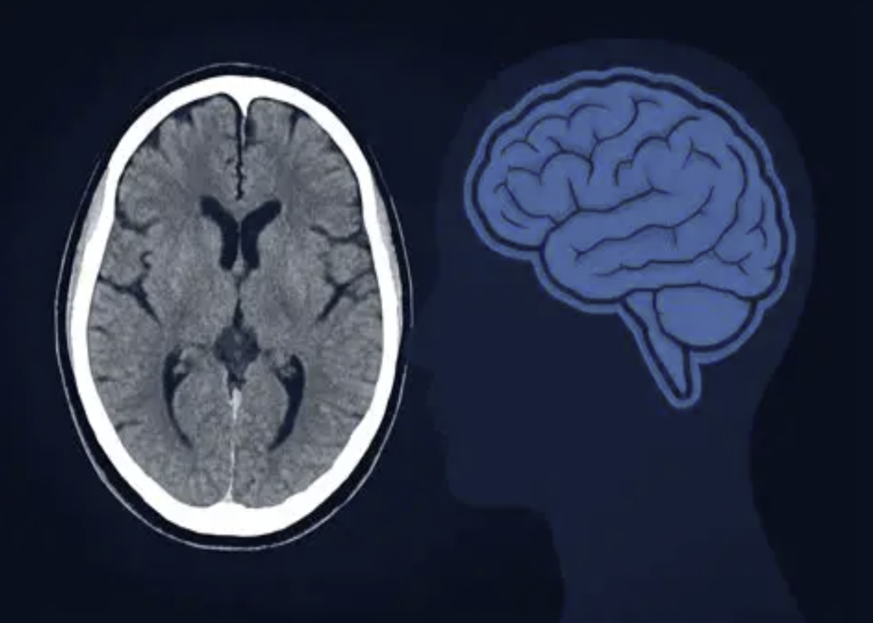
Figure 4. CT brain scan showing anatomical structures. |
The key advantage of CT is its ability to separate overlapping tissues. Structures that would be indistinguishable in a standard X-ray become clearly visible because each slice represents a specific depth within the body. This makes CT especially powerful for detecting tumors, internal bleeding, organ damage, and complex fractures. As a result, CT imaging represents a major milestone in bioimaging, bridging the gap between simple projection imaging and true 3D visualization of human anatomy.
1.5 The Biomedical Imaging Spectrum
Biomedical imaging techniques span a wide range of physical phenomena, and the type of information they provide is strongly determined by where they fall on the electromagnetic spectrum—or, more broadly, the physical spectrum. Different forms of energy interact with biological tissue in different ways, revealing complementary structural, functional, and molecular information. Understanding this spectrum is key to understanding why so many distinct imaging modalities exist.
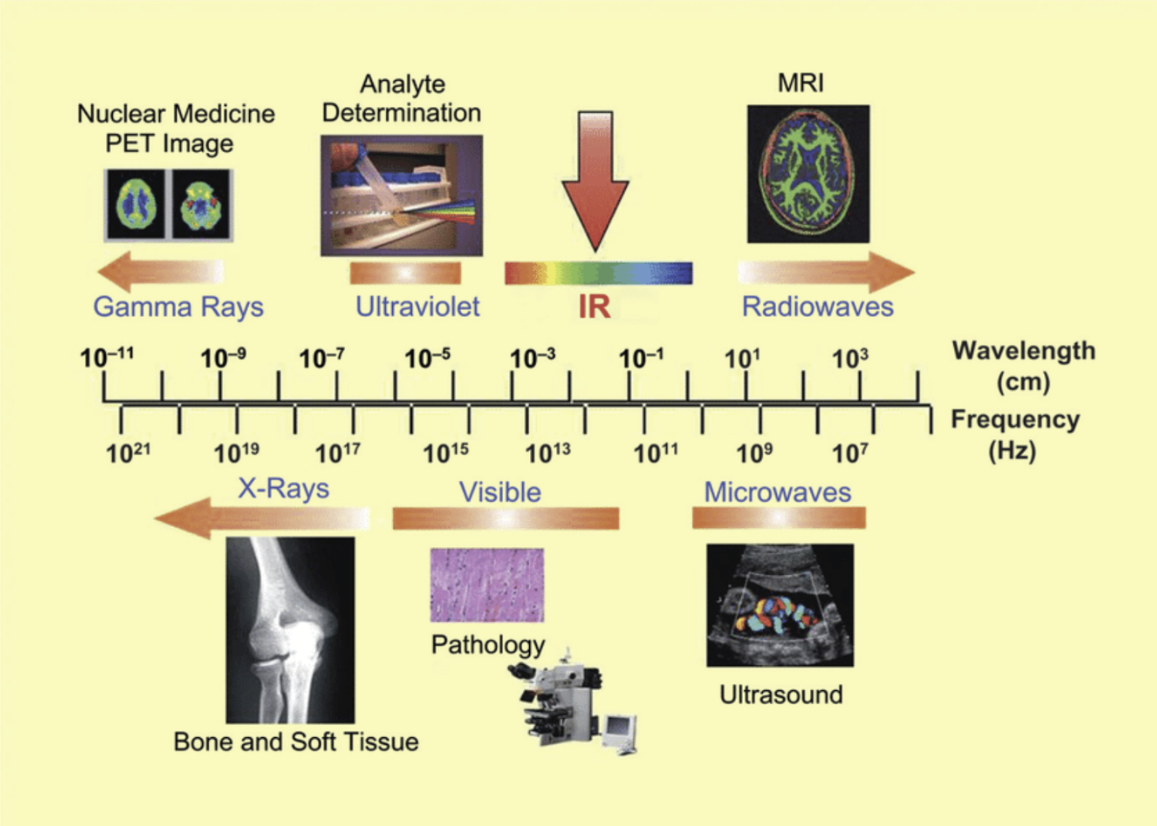
Figure 5. Electromagnetic spectrum showing where different medical imaging modalities fall based on wavelength and frequency. |
At the high-energy end of the spectrum, gamma rays are used in nuclear imaging techniques such as PET and SPECT. These methods do not primarily image anatomy; instead, they visualize physiological and metabolic processes by detecting radioactive tracers inside the body. This makes them especially powerful for studying cancer, brain function, and cardiac health.
Moving to lower energies, radio waves are used in magnetic resonance imaging (MRI). MRI relies on the interaction between radiofrequency signals and nuclear spins in a strong magnetic field, producing highly detailed images of soft tissues. Because it does not use ionizing radiation, MRI is particularly valuable for imaging the brain, muscles, and connective tissues.
In the visible and infrared range, optical imaging techniques are used. These methods can capture fine structural details and molecular signals, often using fluorescence or absorption contrast. Optical imaging is widely used in microscopy, endoscopy, and preclinical research, though light scattering limits how deeply it can penetrate into tissue.
Outside the electromagnetic spectrum altogether, ultrasound imaging uses high-frequency sound waves. By measuring echoes from tissue interfaces, ultrasound provides real-time images of organs, blood flow, and fetal development. It is portable, inexpensive, and safe, making it one of the most commonly used imaging modalities in clinical practice.
No single imaging technique can capture all aspects of biology. As a result, multimodal imaging has become increasingly important. By combining information from multiple imaging modalities—such as PET with CT or MRI—clinicians and researchers can obtain a more complete picture that integrates anatomy, function, and molecular activity. This synergy lies at the heart of modern bioimaging and continues to drive innovation in the field.
1.6 Tomography and Inversion
At the heart of many bioimaging techniques is the idea of tomography, which relies on collecting data from many different angles. Each measurement is a projection—a compressed view of the internal structures along a particular direction. On its own, a single projection contains limited information, much like a shadow that flattens a three-dimensional object into two dimensions.
Tomographic imaging works by acquiring a large number of these projections from different angles around the body or object. The key challenge is inversion: taking the measured projection data and reverse-engineering the internal structure that produced them. In other words, the imaging system asks the question, “What distribution inside the body would give rise to the measurements we observed?” Mathematical inversion algorithms answer this question, allowing the reconstruction of cross-sectional images that reveal internal anatomy without overlapping tissues.
Inversion is not unique to CT; it appears across biomedical imaging, including PET, SPECT, and MRI. While the physics of data acquisition differs between modalities, the unifying concept is the same: indirect measurements are transformed into meaningful images through computation.
1.7 Blurring, Resolution, and Deblurring
No imaging system is perfect. Even when the internal structure is reconstructed correctly, images often appear blurred. This blur arises from physical limitations such as detector size, wave diffraction, motion, and noise. A useful way to describe this effect is through the point spread function (PSF), which characterizes how a single point object appears in the image. Instead of a perfect point, it spreads into a small blob.
Mathematically, image formation can often be modeled as a convolution of the true object with the system’s point spread function. This means the observed image is a blurred version of reality, shaped by the imaging system itself. Understanding this relationship is essential for interpreting images correctly and for improving image quality. Deblurring aims to undo the effects of this convolution and recover a sharper estimate of the original object. This process is closely tied to inversion and often relies on mathematical tools such as the Fourier transform, which provides a powerful framework for analyzing blurring and resolution. These concepts will be developed in detail later, but the key idea is simple: by understanding how blur is introduced, we can design algorithms to partially reverse it.
Together, tomography, inversion, and deblurring form the mathematical backbone of biomedical imaging, turning indirect, imperfect measurements into clear and clinically meaningful images.
1.8 Convolution, Fourier Analysis, and Inversion
A central mathematical idea in biomedical imaging is the relationship between convolution and the Fourier domain. In the spatial or time domain, blurring is commonly modeled as a convolution between the true object and the imaging system’s point spread function. While convolution can be difficult to analyze directly, it becomes much simpler when viewed through the lens of Fourier analysis.
In the Fourier domain, convolution turns into multiplication. This powerful result means that instead of dealing with a complex integral in space or time, we can work with straightforward multiplications in frequency space. This insight underlies many reconstruction and deblurring algorithms in medical imaging. By transforming data into the Fourier domain, correcting or compensating for system effects, and transforming back, imaging systems can recover clearer and more accurate representations of internal structures.
These ideas connect directly to inversion. Inversion starts with the outcome—the measured image or signal—and infers the underlying cause: the original biological structure or process. Whether reconstructing a CT slice from projections or deblurring an optical image, inversion reframes imaging as a problem of reasoning backward from data to source. This perspective is a unifying theme across nearly all biomedical imaging modalities.
1.9 Biomedical Imaging as an Interdisciplinary
Medical imaging is inherently interdisciplinary, sitting at the intersection of multiple scientific and technical domains. Mathematics provides the language of inversion, reconstruction, and signal processing. Physics governs how energy—whether X-rays, radio waves, light, or sound—interacts with tissue. Chemistry enables contrast agents and molecular probes. Biology explains the structure and function of tissues being imaged, while medicine defines the clinical questions that imaging must answer. Engineering brings all of these elements together into practical, reliable imaging systems.
This convergence is the foundation of biomedical engineering, a field that explicitly integrates biology and medicine with engineering principles. As healthcare becomes increasingly technology-driven—relying on advanced imaging, data analysis, and computational tools—the demand for biomedical engineers continues to grow. Biomedical imaging exemplifies this trend, offering opportunities to work at the cutting edge of science while directly impacting patient care.
1.10 X-Ray Imaging, the Radon Transform, and Fourier-Based Reconstruction
X-ray imaging begins with a line integral model. As X-rays pass through an object, they are attenuated by the material along their path. The detector measures the energy (or intensity) of the transmitted X-ray photons, and this measurement corresponds mathematically to an integral of the object’s attenuation properties along the X-ray path. Each measurement therefore captures cumulative information along a straight line through the object.
When this process is repeated across many parallel rays, the result is a collection of line integrals that form the Radon transform of the object. If the object is represented as a two-dimensional function
\[f(x, y)\]then the measured data can be described as a new function
\[p(t, \theta).\]Here, $t$ represents position along a one-dimensional detector array, and $\theta$ represents the orientation angle of the X-ray projection. By changing the angle $\theta$, the data acquisition system rotates around the object and gathers projections from many different directions.
The central goal of X-ray computed tomography is to reconstruct the original image
\[f(x, y)\]from the measured projection data
\[p(t, \theta).\]This reconstruction problem is an example of inversion: starting with indirect measurements and inferring the internal structure that produced them. The key mathematical tool that makes this possible is Fourier analysis.
For each fixed angle $\theta$, a one-dimensional Fourier transform is applied to the corresponding projection
\[p(t, \theta).\]A remarkable result—known as the Fourier slice theorem—states that the Fourier transform of a projection corresponds to a slice through the two-dimensional Fourier transform of the original image. Each 1D projection therefore provides a line (or profile) of information in the frequency domain.
As the acquisition system rotates and projections are collected over many angles, these frequency-domain slices fill up the entire two-dimensional Fourier space of the object. Once sufficient coverage of this Fourier space is obtained, the original image can be reconstructed by applying the inverse Fourier transform. This framework highlights a deep connection between data acquisition and image reconstruction. The forward process—collecting X-ray projections as line integrals—is mathematically linked to the inverse process—reconstructing the image—through Fourier analysis. Understanding this relationship is fundamental to biomedical imaging, as it reveals how physical measurements, mathematical transforms, and computational algorithms work together to produce meaningful images of the human body.
1.11 Positron Emission Tomography (PET): Imaging Function, Not Just Structure
Positron emission tomography (PET) is a medical imaging technique designed to visualize functional activity inside the body rather than detailed anatomy. Instead of showing what organs look like, PET reveals how tissues are behaving—such as how actively they are consuming energy or participating in metabolic processes. This makes PET especially powerful for studying cancer, brain function, and heart disease.
PET imaging begins by introducing a radioactive tracer into the body. These tracers are radioactive chemicals that are biologically active and participate in normal physiological processes. For example, many PET tracers are glucose analogs. Because tumors often have abnormally high metabolic rates, they consume more glucose than surrounding tissue and therefore absorb more of the tracer. This selective uptake creates contrast based on biological function rather than physical density. The tracer undergoes radioactive decay by emitting a positron. When the positron encounters an electron, the two annihilate, producing a pair of gamma-ray photons. These two photons are emitted simultaneously and travel in nearly opposite directions. Gamma rays, like X-rays, can penetrate tissue and escape the body, allowing them to be detected externally.
PET scanners are designed with rings of detectors surrounding the patient. When two detectors on opposite sides of the scanner register gamma-ray photons at the same time, the system infers that both photons originated from the same annihilation event. This event must have occurred somewhere along the straight line connecting the two detectors, known as a line of response. By collecting millions of these coincident events from many angles, PET builds up a dataset of projection measurements.
As with CT, image formation in PET is fundamentally an inversion problem. The measured projection data are processed using mathematical reconstruction techniques—many of which rely on Fourier analysis—to reconstruct a three-dimensional image of tracer concentration inside the body. The resulting image maps metabolic or biochemical activity across tissues.
A key strength of PET is its complementarity with CT. CT provides high-resolution anatomical detail, clearly showing the shapes and boundaries of organs, while PET reveals biological function, such as metabolism occurring within those structures. Together, they allow clinicians to see not only where structures are, but what they are doing, making PET a cornerstone of functional bioimaging.
1.12 Functional vs. Anatomical Imaging
Medical imaging techniques can be broadly divided into anatomical imaging and functional imaging, based on the type of information they provide. This distinction is central to understanding why multiple imaging modalities are often used together in clinical practice.
Computed Tomography (CT) is a classic example of anatomical imaging. It provides highly detailed images of the body’s internal structure, clearly showing the size, shape, and spatial relationships of organs, bones, and tissues. CT excels at answering questions such as Where is the tumor?, Is there a fracture?, or What is the shape of this organ? However, it does not directly reveal how tissues are functioning.
In contrast, nuclear imaging techniques, such as PET and SPECT, are forms of functional imaging. These modalities focus on physiological and biological activity rather than structure. By tracking radioactive tracers that participate in metabolic or biochemical processes, nuclear imaging shows how tissues are functioning—such as how much glucose a tumor is consuming or how lood flows through the heart or brain.
The key goal of functional imaging is to understand where biological activity is occurring in the body and how much activity is present. Instead of visualizing anatomy alone, functional imaging maps processes like metabolism, perfusion, and receptor binding. When combined with anatomical imaging, this functional information provides a much more complete picture of health and disease, linking structure to function in a way that is essential for modern bioimaging.
1.13 Positron Emission Tomography (PET)
Positron Emission Tomography (PET) is a powerful imaging technique that visualizes biological function rather than merely anatomical structure. PET is widely used to study physiological processes across the body, providing insight into metabolism, cancer activity, brain function, and heart conditions. For example, PET can track how tissues consume energy or nutrients, detect tumors based on their increased metabolic activity, map regions of the brain involved in cognition or disease, and assess blood flow and tissue viability in the heart. By capturing dynamic biological processes, PET complements anatomical imaging techniques such as CT and MRI.
PET imaging relies on the administration of radiotracers, which are biologically active molecules labeled with a radioactive isotope. These tracers participate in normal metabolic processes and undergo decay that emits paired gamma-ray photons. The photons travel in opposite directions, and PET scanners detect coincident events, where two photons are simultaneously recorded by detectors on opposite sides of the body. This coincidence detection provides precise spatial information about the location of the radioactive decay event, allowing for accurate mapping of biological activity within tissues.
The signals collected by the PET scanner are processed using computational algorithms to generate tomographic three-dimensional images. These images display the distribution and intensity of the tracer, effectively revealing where and how tissues are functioning in real time. Unlike CT, which primarily provides structural information, PET highlights active physiology, making it an indispensable tool in clinical applications such as oncology, neurology, and cardiology. By combining functional insight with high-resolution imaging, PET plays a critical role in diagnosis, treatment planning, and monitoring of disease progression.
1.14 Single Photon Emission Computed Tomography (SPECT)
Single Photon Emission Computed Tomography (SPECT) is a nuclear imaging technique designed to visualize functional activity within the body, much like PET, but it differs in how radioactive decay is detected. In SPECT, the radiotracer emits single, unpaired gamma-ray photons. These photons are emitted randomly and individually, and a collimator is used to ensure that only photons traveling along specific directions reach the detector. Each detected photon provides information about its path through the body, which contributes to the overall dataset used for image reconstruction.
The reconstruction process in SPECT involves collecting projection images from multiple angles around the patient. Unlike PET, which benefits from coincident paired photon detection, SPECT requires more sophisticated preprocessing to account for the single-photon nature of the data. Computational algorithms then combine these projections to generate three-dimensional images of radiotracer activity, effectively mapping physiological processes such as blood flow, metabolism, or organ function.
Although SPECT generally has lower spatial resolution compared to PET due to the single-photon detection mechanism, it remains a valuable tool for functional imaging. Both SPECT and PET produce tomographic images that reveal the distribution of radiotracers inside the body. These images allow clinicians to observe physiological activity that cannot be captured by purely anatomical imaging methods, making SPECT an important modality for diagnosing and monitoring disease.
1.15 Magnetic Resonance Imaging (MRI)
Magnetic Resonance Imaging (MRI) is fundamentally different from X-ray and nuclear imaging because it does not rely on ionizing radiation. Instead, MRI uses strong magnetic fields and radio-frequency (RF) signals to probe the body’s internal structures. When a patient is placed in a strong magnetic field, the hydrogen nuclei (protons) in the body align with the field. An RF pulse is then applied to disrupt this alignment, and as the protons relax back to their original orientation, they emit RF signals that are measured by the scanner. These signals are processed using computational algorithms to generate detailed images of internal anatomy.
MRI is particularly powerful because it is highly sensitive to water content and the micro-environment of tissues. This sensitivity makes MRI excellent for imaging soft tissues such as the brain, muscles, and internal organs. Beyond structural imaging, MRI can detect chemical shifts in different molecular environments, identify tumors and inflammation, and observe functional activity indirectly through blood flow changes in the brain. This combination of structural and functional information allows MRI to bridge the gap between anatomical and physiological imaging.
Unlike CT, which is optimized for detailed structural imaging, or PET/SPECT, which primarily visualizes functional processes, MRI provides both high-resolution anatomical images and functional information in a single modality. Its ability to distinguish soft tissues with exquisite contrast, coupled with the absence of ionizing radiation, makes MRI a cornerstone of modern biomedical imaging. Today, MRI is widely used in neurology, cardiology, and musculoskeletal medicine, offering unparalleled insight into both the structure and function of tissues in the human body.
1.16 Ultrasound Imaging
Ultrasound imaging is one of the most widely used medical imaging techniques due to its affordability, safety, speed, and versatility. It has become increasingly important in point-of-care imaging, where clinicians need rapid, real-time information at the bedside or in outpatient settings. Unlike X-ray, CT, or nuclear imaging, ultrasound uses mechanical sound waves rather than electromagnetic radiation to visualize internal structures.
The imaging process relies on a transducer that contains piezoelectric materials. When an electrical signal is applied, the transducer generates high-frequency sound waves that propagate into the body. These waves reflect off tissues at interfaces where acoustic properties change, and the returning echoes are detected by the same transducer. Sophisticated algorithms then reconstruct these reflections into two- or three-dimensional images, producing a real-time view of internal anatomy.
Ultrasound offers several key advantages. It is completely non-invasive and does not expose patients to ionizing radiation, making it safe for repeated use, including in fetal imaging. Ultrasound imaging is also highly cost-effective, portable, and capable of providing real-time images, which is critical for applications such as monitoring blood flow, guiding biopsies, or assessing organ motion. Despite its versatility, ultrasound has limitations, including lower spatial resolution and soft tissue contrast compared to MRI or CT.
Looking toward the future, AI-enhanced ultrasound holds significant potential. Machine learning algorithms can assist with image interpretation, automate measurements, and improve diagnostic accuracy, making ultrasound even more valuable as a frontline imaging modality in modern healthcare. Its combination of safety, speed, and real-time capability ensures that ultrasound will remain a core tool in both clinical and point-of-care settings.
1.17 Optical Imaging
Optical imaging is a biomedical imaging technique that uses visible and infrared light to probe biological tissues. Unlike X-ray or nuclear imaging, which provide structural or metabolic information, optical imaging focuses on molecular and cellular-level interactions, enabling researchers and clinicians to study biological processes at a microscopic scale. Its unique strengths include the ability to extract specific biological signatures and observe fine details that are otherwise invisible to traditional imaging methods.
One important form of optical imaging is luminescence imaging, where proteins, genes, or other cellular targets are tagged with luminescent probes. These tagged targets emit light naturally, a process known as bioluminescence, allowing for passive imaging because the light is produced by the body itself rather than an external source. To localize the sources of luminescence inside the body, bioluminescence tomography (BLT) collects emitted light from multiple external views. This data is combined with a tomographic anatomical reference—often from CT or MRI—to solve a forward model that predicts the expected signals and an inverse problem that reconstructs the internal distribution of light-emitting sources.
Optical imaging faces significant challenges, particularly in the inverse reconstruction, because light scatters and absorbs as it travels through tissue, making it difficult to accurately determine the origin of the signal. Other optical techniques extend the capabilities of this modality. Fluorescence tomography uses externally excited light to image specific molecular targets, while Optical Coherence Tomography (OCT) provides high-resolution structural imaging, commonly used in retinal imaging. Together, these techniques allow optical imaging to provide both functional and structural information at scales ranging from single cells to whole organs, offering a complementary perspective to other biomedical imaging modalities.
1.18 Image Processing and Analysis
Acquiring an image is only the first step in biomedical imaging; image processing and analysis are essential to extract meaningful information. Raw images often contain noise, overlapping structures, or low contrast, making it difficult to interpret them directly. By applying computational techniques, clinicians and researchers can enhance image quality, isolate regions of interest, and quantify biological features.
One key aspect of image analysis is segmentation, which involves isolating specific organs, tumors, blood vessels, or other anatomical structures from surrounding tissue. Segmentation enables more precise measurements, such as tumor volume or vessel diameter, and serves as the foundation for further analyses. Classification is another important technique, in which tissues or structures are categorized based on their characteristics, helping to distinguish healthy from diseased tissue or identify specific functional regions.
Enhancement techniques improve image quality for better interpretation. Methods such as deblurring reverse the effects of the imaging system’s point spread function, while contrast improvement increases the visibility of subtle features. Visualization tools also play a critical role, including emerging technologies like Augmented Reality (AR), which can overlay imaging data onto the patient during surgery, enabling AR-assisted image-guided procedures. While AR is not the primary focus here, it illustrates the growing integration of imaging with clinical practice.
In this course, the focus is on bioimaging and bioinstrumentation, with a particular emphasis on tomographic imaging. By understanding both the physics of image formation and the computational tools used for reconstruction and analysis, students gain the skills needed to transform raw imaging data into clinically and scientifically meaningful insights.
1.19 Phenotype vs. Genotype
In biology, it is important to distinguish between genotype and phenotype. The genotype refers to an organism’s genetic makeup, encoded in its DNA, and represents the blueprint of potential traits and biological processes. In contrast, the phenotype encompasses the observable traits, functions, and disease states that result from the interaction of the genotype with the environment. Phenotype includes characteristics such as organ structure, metabolic activity, and physiological responses, all of which can be studied using biomedical imaging.
Biomedical imaging is primarily concerned with the phenotype, providing detailed information about tissue structure, function, and behavior in living organisms. Techniques such as CT, MRI, PET, and optical imaging reveal how cells, organs, and systems are functioning, enabling clinicians to observe the physical manifestation of disease or biological processes. Meanwhile, bioinformatics and genetic analysis provide complementary genotype data, identifying the underlying DNA sequences and genetic variants that may influence these traits.
A major goal in modern biomedical research is to link genotype to phenotype, bridging the gap between genetic information and observable outcomes. Understanding this relationship forms the foundation for personalized medicine, where treatments and interventions are tailored to an individual’s genetic profile and physiological characteristics. By integrating imaging-based phenotypic data with genetic insights, clinicians can achieve better diagnosis, targeted therapies, and more accurate predictions of disease progression and treatment response.
1.20 AI, Machine Learning, and the Future of Imaging
Medical imaging is a rapidly evolving field, and its growth is being accelerated by advances in artificial intelligence (AI) and machine learning, particularly deep learning. These technologies offer new ways to process, analyze, and interpret imaging data, enabling improvements that were previously impractical or impossible. AI can enhance image reconstruction, reduce noise and artifacts, and generate higher-quality images from lower doses of radiation or faster acquisition times.
Beyond reconstruction, AI is increasingly applied to automated analysis. Machine learning algorithms can segment tissues, classify abnormalities, and detect subtle patterns that may be difficult for humans to identify. AI also supports enhanced interpretation, providing clinicians with quantitative metrics, predictive models, and decision-support tools that improve accuracy and efficiency. This combination of AI and imaging is transforming both research and clinical practice.
Looking ahead, the future of biomedical imaging may include AI-assisted or even AI-led image interpretation, where algorithms guide diagnostic decisions, highlight areas of concern, and integrate data from multiple modalities. Robotics guided by multimodal imaging is also emerging, enabling more precise surgeries and interventions. Importantly, the goal of these technologies is to enhance healthcare, not replace medical professionals. For students, AI in imaging presents opportunities to engage in real-world research projects, combining imaging and machine learning to gain hands-on experience and contribute to the next generation of biomedical innovation.